
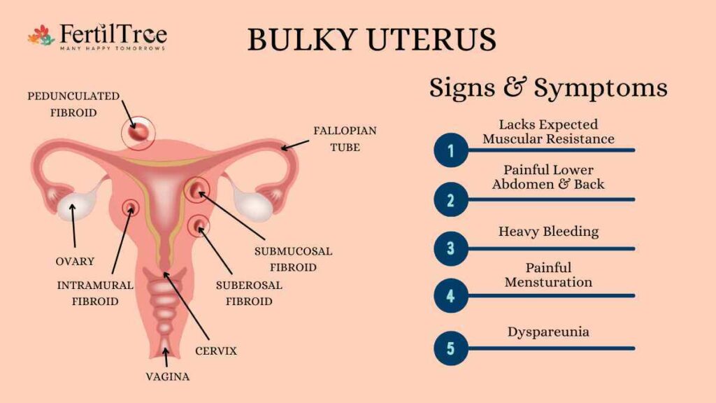
Bulky Uterus: Symptoms, Causes, and Treatment

Bulky Uterus: Symptoms, Causes, and Treatment

A bulky uterus isn’t a diagnosis on its own — it’s a description, and what it means for you depends entirely on what’s causing it. A uterus that’s mildly enlarged from a past pregnancy is a very different situation from one enlarged by diffuse adenomyosis, even though both can show up as the same word on your ultrasound report. This guide breaks down what actually changes the answer — the cause behind the finding, not the finding itself — including real IVF-outcome data most bulky-uterus content leaves out entirely.

Key takeaways

●       “Bulky uterus” describes a finding, not a cause — fibroids, adenomyosis, and physiological enlargement carry very different fertility implications.

●       Adenomyosis has the clearest documented IVF impact: one study found ongoing pregnancy rates of 11.1% with adenomyosis vs. 45.9% without it.

●       The number of adenomyosis features on ultrasound matters — pregnancy chances decline in a dose-response pattern as more features are present.

●       How your fibroids are positioned matters more than whether you have them — cavity-distorting fibroids carry more risk than small intramural ones.

●       GnRH-agonist pretreatment before transfer may help in diffuse adenomyosis specifically — a treatment-sequencing detail generic content rarely covers.

●       Your surgeon’s read of your specific scan always outweighs any general guide, including this one.

Why the Cause Behind Your Bulky Uterus Changes the Answer?

“Bulky uterus” is a radiology description, not a diagnosis — it just means your uterus measured larger than the typical range on a scan. Some enlargement is completely normal; a uterus that’s carried a pregnancy is often mildly larger for life, with no fertility implications at all. Other times the same word on a report points to fibroids, adenomyosis, or occasionally something that needs prompt attention like pyometra or endometrial changes. Treating every bulky-uterus finding as equally significant — the way a lot of generic content does — misses the fact that a physiologically enlarged uterus and a uterus with diffuse adenomyosis are, for fertility purposes, almost entirely different conversations.

Why the Cause Behind Your Bulky Uterus Changes the Answer?

A non-pregnant uterus typically measures about 7–8 cm long, 5 cm wide, and 4 cm thick, with a volume of roughly 80–200 mL. Beyond that range, here’s how the leading causes typically compare for fertility purposes:

Cause

Typical Fertility Impact

What the Evidence Shows

Physiological (past pregnancy)

Minimal to none

No consistent link to reduced fertility on its own

Small intramural / subserosal fibroids

Usually minimal

Small fibroids not distorting the cavity often have little measurable effect

Submucosal or cavity-distorting fibroids

Meaningful

Associated with reduced implantation; typically warrant evaluation before IVF

Diffuse adenomyosis

Significant

Ongoing pregnancy rate 11.1% vs. 45.9% without it in one study (see chart below)

This is a general guide to typical patterns, not a prediction for any individual case — your own imaging and history determine what applies to you.

Bulky Uterus Self-Check

Beyond the ultrasound report itself, how you’re feeling day to day is a reasonable first filter for how soon to act.

How You’re Feeling

Likely Next Step

Found incidentally on a routine scan, no symptoms, periods normal

Reasonable to discuss at your next routine visit, not urgent

Heavier or longer periods, some pelvic pressure, otherwise stable

Worth a dedicated conversation with your gynaecologist soon

Heavy bleeding with fatigue or anemia symptoms, trying to conceive 6–12+ months, or any postmenopausal bleeding

See a specialist promptly — don’t wait for your next routine visit

This is a general guide, not a diagnosis — postmenopausal bleeding in particular should always be evaluated promptly regardless of how mild it seems.

What Most Bulky Uterus Content Skips?

Most bulky-uterus articles will tell you adenomyosis “can affect fertility.” Very few show you the actual numbers, or the fact that severity matters, not just presence or absence.

Ongoing pregnancy rate after IVF, with vs. without adenomyosis.

A second, less-cited finding matters just as much: the effect isn’t all-or-nothing. A prospective UCL study scored adenomyosis by the number of features visible on 3D ultrasound (out of seven possible) and found a clear dose-response relationship — pregnancy chances fell step by step as more features were present, independent of age and ovarian reserve.

Clinical pregnancy rate by number of adenomyosis ultrasound features present.

There’s a treatment-sequencing detail that follows from this, and it’s genuinely rare in patient-facing content: several studies have found that pretreating diffuse adenomyosis with a GnRH agonist before a frozen embryo transfer — an “ultra-long” downregulation protocol rather than the standard long protocol — is associated with meaningfully better outcomes (one study reported clinical pregnancy rates of 55.3% vs. 37.9%, and live birth rates of 43.4% vs. 25.9%, favoring the longer downregulation approach in diffuse adenomyosis specifically). Separately, more recent research has explored oxytocin-receptor antagonists given around the time of transfer to calm excess uterine contractions in adenomyosis, with early studies reporting a reduction in early miscarriage. Neither is a guarantee, and both are protocol decisions for your treating specialist — but they’re worth knowing exist before you assume a bulky uterus finding rules out IVF success.

Reading Your Ultrasound Report

A few terms that commonly appear on a pelvic ultrasound report, decoded:

  • Uterine volume (mL) — the calculated size of the uterus; above roughly 200 mL in a non-pregnant woman is generally what prompts the “bulky” description.
  • AP diameter — the front-to-back measurement of the uterus in centimetres; radiologists often reference this alongside length and width.
  • Junctional zone thickening — a specific sign radiologists look for at the border between the uterine lining and muscle; this is one of the clearest ultrasound markers of adenomyosis specifically, not fibroids.
  • “Size equivalent to X weeks of pregnancy” — a common shorthand doctors use to describe uterine size using pregnancy-week terms as a size reference, not an indication that anything pregnancy-related is happening.

None of these terms alone tell you what to do next — that’s genuinely your specialist’s call — but knowing what they mean makes that conversation easier to follow.

What Happens Next?

  • Diagnosis — usually a pelvic exam plus transvaginal ultrasound; MRI is added when more detail is needed, particularly to tell adenomyosis and fibroids apart or map fibroid location before surgery. Our page on diagnosis of infertility covers the broader work-up.
  • Treatment options — range from hormonal medication and GnRH agonists to myomectomy for fibroids that are affecting the cavity, up to hysterectomy for significant, symptomatic cases once childbearing is complete. Our infertility management page and, where IVF is the right next step, our team can map a protocol to your specific scan findings.
  • Where this fits regionally — fibroids are especially common across South Asia, including India, where Global Burden of Disease data shows one of the steeper rises in prevalence over the past three decades — worth knowing if you’re weighing how much to read into a single finding.

If a related condition on a scan report has you searching for answers too, our guide on thin endometrium lining covers the other uterine-factor finding we’re asked about most — the two sometimes share an underlying cause. You can also get in touch with our clinic directly to talk through your own scan results.


Bottom line

“Bulky uterus” is a description, not a verdict — what it means for your fertility depends on the cause, and for adenomyosis specifically, on how much of it is present. Real data exists on both points; most bulky-uterus content simply doesn’t show it to you.

Want your specific scan talked through rather than matched against a generic checklist? Book a consultation at FertilTree — we can walk through your actual results and what they mean for your next step.


Frequently Asked Questions

Can I get pregnant with a bulky uterus?

Often, yes — especially with mild or physiological enlargement, or small fibroids. Diffuse adenomyosis or cavity-distorting fibroids are more likely to need evaluation first.

Does adenomyosis definitely mean IVF won't work?

No — it lowers the odds on average, and more so with more ultrasound features present, but plenty of women with adenomyosis go on to have successful pregnancies, sometimes with adjusted protocols like GnRH-agonist pretreatment.

What is a normal uterus size?

Roughly 7–8 cm long, 5 cm wide, and 4 cm thick, with a volume of about 80–200 mL in a non-pregnant woman.

What does “junctional zone thickening” mean on my report?

It’s a specific ultrasound sign radiologists use to identify adenomyosis, distinct from fibroids — worth asking your doctor about directly if it appears on your report.

Should an enlarged uterus be removed?

Not usually for mild cases. Hysterectomy is generally reserved for significant, symptomatic fibroids or adenomyosis once childbearing is complete; conservative treatment is preferred otherwise.

How common are uterine fibroids?

They affect roughly 20–30% of women of reproductive age, rising to a cumulative incidence of about 70–80% by age 50.

MEDICALLY REVIEWED BY

Dr. Firuza Parikh

MD, PhD, Director, Jaslok-FertilTree International Fertility Centre  ·  Last updated August 2026

Uterine-factor findings — bulky uterus, adenomyosis, fibroids — are among the most common reasons patients come to Dr. Parikh for a second opinion before starting or continuing IVF, and the conversation almost always turns on the specific cause and severity rather than the finding itself. She trained in IVF and Reproductive Endocrinology at Yale University School of Medicine under Dr. Alan DeCherney, established one of India’s first private-hospital IVF centres at Jaslok Hospital in 1989, and delivered South-East Asia’s first ICSI baby in 1994. Over more than three decades in reproductive medicine, she and her team have helped deliver more than 20,000 babies for patients from over 60 countries. Read more at Dr. Firuza Parikh’s full profile.